Friday, September 25, 2015

Dunaliella salina - a micro alga producing Beta -carotene


          Dunaliella salina is a type of halophile green micro algae especially found in sea salt fields. Known for its antioxidant activity because of its ability to create large amount of carotenoids, it is used in cosmetics and dietary supplements. Few organisms can survive in such highly saline conditions as salt evaporation ponds. To survive, these organisms have high concentrations of β-carotene to protect against the intense light, and high concentrations of glycerol to provide protection against osmotic pressure. This offers an opportunity for commercial biological production of these substances.

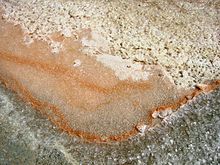
          From a first pilot plant for Dunaliella cultivation for β-carotene production established in the USSR in 1966, the commercial cultivation of Dunaliella for the production of β-carotene throughout the world is now one of the success stories of halophile biotechnology. Different technologies are used, from low-tech extensive cultivation in lagoons to intensive cultivation at high cell densities under carefully controlled conditions. Although Dunaliella salina produce β-carotene in a high salt environment,Archaea such as Halobacterium, not Dunaliella, are responsible for the red and pink coloring of salt lakes.[1] Occasionally, orange patches of Dunaliella colonies 
will crop up.
        Attempts have been made to exploit the high concentrations of glycerol accumulated by
Dunaliella as the basis for the commercial production of this compound. Although technically the production of glycerol from Dunaliella was shown to be possible, economic feasibility is low and no biotechnological operation exists to exploit the alga for glycerol production.
Dunaliella salina
FleurDeSel.JPG
Orange-colored Dunaliella salina within sea salt
Scientific classification
Kingdom:Plantae
Phylum:Chlorophyta
Class:Chlorophyceae
Order:Volvocales
Family:Dunaliellaceae
Genus:Dunaliella
Species:D. salina
Binomial name
Dunaliella salina

Reference : 

Chlamydomonas reinhardtii - A model Unicellular Alga for Biology


Chlamydomonas reinhardtii
Chlamydomonas6-1.jpg
Scientific classification
Kingdom:Plantae
Phylum:Chlorophyta
Class:Chlorophyceae
Order:Chlamydomonadales
Family:Chlamydomonadaceae
Genus:Chlamydomonas
Species:C. reinhardtii
Binomial name
Chlamydomonas reinhardtii
P.A.Dang.
Chlamydomonas reinhardtii is a single-cell green alga about 10 micrometres in diameter that swims with twoflagella. It has a cell wall made of hydroxyproline-rich glycoproteins, a large cup-shaped chloroplast, a largepyrenoid, and an "eyespot" that senses light.
Chlamydomonas species are widely distributed worldwide in soil and fresh water. Chlamydomonas reinhardtii is an especially well studied biological model organism, partly due to its ease of culturing and the ability to manipulate its genetics. When illuminated, C. reinhardtii can grow photoautotrophically, but it can also grow in the dark if supplied with organic carbon. Commercially, C. reinhardtii is of interest for producing biopharmaceuticals and biofuel, as well being a valuable research tool in making hydrogen.

History

The C. reinhardtii wild-type laboratory strain c137 (mt+) originates from an isolate made near Amherst, Massachusetts, in 1945 by Gilbert M. Smith.
The species has been spelled several different ways because of different transliterations of the name from Russian: reinhardireinhardii, and reinhardtii all refer to the same species, C. reinhardtii Dangeard.

Model organism

Cross section of a Chlamydomonas reinhardtii algae cell, a 3D representation
Chlamydomonas is used as a model organism for research on fundamental questions in cell and molecular biology such as:
  • How do cells move?
  • How do cells respond to light?
  • How do cells recognize one another?
  • How do cells generate regular, repeatable flagellar waveforms?
  • How do cells regulate their proteome to control flagellar length?
  • How do cells respond to changes in mineral nutrition? (nitrogen, sulfur, etc.)
There are many known mutants of C. reinhardtii. These mutants are useful tools for studying a variety of biological processes, including flagellar motility, photosynthesis, and protein synthesis. Since Chlamydomonasspecies are normally haploid, the effects of mutations are seen immediately without further crosses.
In 2007, the complete nuclear genome sequence of C. reinhardtii was published.[4]
Channelrhodopsin-1 and Channelrhodopsin-2, proteins that function as light-gated cation channels, were originally isolated from C. reinhardtii.These proteins and others like them are increasingly widely used in the field of optogenetics.

Reproduction

Vegetative cells of the reinhardtii species are haploid with 17 small chromosomes. Under nitrogen starvation, vegetative cells differentiate into haploid gametes.There are two mating types, identical in appearance and known as mt(+) and mt(-), which can fuse to form a diploid zygote. The zygote is not flagellated, and it serves as a dormant form of the species in the soil. In the light, the zygote undergoes meiosis and releases four flagellated haploid cells that resume the vegetative lifecycle.
Under ideal growth conditions, cells may sometimes undergo two or three rounds of mitosis before the daughter cells are released from the old cell wall into the medium. Thus, a single growth step may result in 4 or 8 daughter cells per mother cell.
The cell cycle of this unicellular green algae can be synchronized by alternating periods of light and dark. The growth phase is dependent on light, whereas, after a point designated as the transition or commitment point, processes are light-independent.

Genetics

The attractiveness of the alga as a model organism has recently increased with the release of several genomic resources to the public domain. The Chlre3 draft of the Chlamydomonas nuclear genome sequence prepared by Joint Genome Institute of the U.S. Dept of Energy comprises 1557 scaffolds totaling 120 Mb. Roughly half of the genome is contained in 24 scaffolds all at least 1.6 Mb in length. The current assembly of the nuclear genome is available online.
The ~15.8 Kb mitochondrial genome (database accession: NC_ 001638) is available online at the NCBI database. The complete >200 Kb chloroplast genome is available online.
In addition to genomic sequence data, there is a large supply of expression sequence data available as cDNA libraries and expressed sequence tags (ESTs). Seven cDNA libraries are available online. A BAC library can be purchased from the Clemson University Genomics Institute. There are also two databases of >50 000 and >160 000 ESTs available online.

Evolution

Chlamydomonas has been used to study different aspects of evolutionary biology and ecology. It is an organism of choice for many selection experiments because (1) it has a short generation time, (2) it is both a heterotroph and a facultative autotroph, (3) it can reproduce both sexually and asexually, and (4) there is a wealth of genetic information already available.
Some examples (nonexhaustive) of evolutionary work done with Chlamydomonas include the evolution of sexual reproduction, the fitness effect of mutations,and the effect of adaptation to different levels of CO2.
According to one frequently cited theoretical hypothesis, sexual reproduction (in contrast to asexual reproduction) is adaptively maintained in benign environments because it reduces mutational load by combining deleterious mutations from different lines of descent and increases mean fitness. However, in a long-term experimental study of C. reinhardtii, evidence was obtained that contradicted this hypothesis. In sexual populations, mutation clearance was not found to occur and fitness was not found to increase.

DNA transformation techniques

Gene transformation occurs mainly by homologous recombination in the chloroplast and heterologous recombination in the nucleus. The C. reinhardtii chloroplast genome can be transformed using microprojectile particle bombardment or glass bead agitation, however this last method is far less efficient. The nuclear genome has been transformed with both glass bead agitation and electroporation. The biolistic procedure appears to be the most efficient way of introducing DNA into the chloroplast genome. This is probably because the chloroplast occupies over half of the volume of the cell providing the microprojectile with a large target. Electroporation has been shown to be the most efficient way of introducing DNA into the nuclear genome with maximum transformation frequencies two orders of magnitude higher than obtained using glass bead method.

Production of biopharmaceuticals

Genetically engineered Chlamydomonas reinhardtii has been used to produce a mammalian serum amyloid protein, a human antibody protein, human Vascular endothelial growth factor, a potential therapeutic Human Papilloma virus 16 vaccine,a potential malaria vaccine, and a complex designer drug that could be used to treat cancer.

Clean source of hydrogen production

In 1939, the German researcher Hans Gaffron (1902–1979), who was at that time attached to the University of Chicago, discovered the hydrogen metabolism of unicellular green algae. Chlamydomonas reinhardtii and some other green algae can, under specified circumstances, stop producing oxygen and convert instead to the production of hydrogen. This reaction by hydrogenase, an enzyme active only in the absence of oxygen, is short-lived. Over the next thirty years, Gaffron and his team worked out the basic mechanics of this photosynthetic hydrogen production by algae.
To increase the production of hydrogen, several tracks are being followed by the researchers.
  • The first track is decoupling hydrogenase from photosynthesis. This way, oxygen accumulation can no longer inhibit the production of hydrogen. And, if one goes one step further by changing the structure of the enzyme hydrogenase, it becomes possible to render hydrogenase insensitive to oxygen. This makes a continuous production of hydrogen possible. In this case, the flux of electrons needed for this production no longer comes from the production of sugars but is drawn from the breakdown of its own stock of starch.
  • A second track is to interrupt temporarily, through genetic manipulation of hydrogenase, the photosynthesis process. This inhibits oxygen's reaching a level where it is able to stop the production of hydrogen.
  • The third track, mainly investigated by researchers in the 1950s, is chemical or mechanical methods of removal of O2 produced by the photosynthetic activity of the algal cells. These have included the addition of O2 scavengers, the use of added reductants, and purging the cultures with inert gases. However, these methods are not inherently scalable, and may not be applicable to applied systems. New research has appeared on the subject of removing oxygen from algae cultures, and may eliminate scaling problems.
  • A fourth track has been investigated, namely using copper salts to decouple hydrogenase action from oxygen production.[28]


Sapria himalayana (Rafflesiaceae) from Mizoram

         Griffith (1844) established the genus Sapria with a species S. himalayana (Rafflesiaceae) from Mishmi Hills of erstwhile Assam, now in Arunachal Pradesh. This genus consists of three species, viz., S. himalayana Griff., S. poilanei Gagnep. and S. ram Banziger & B. Hansen and is confined to Southeast Asia with restricted disjunctive distribution.

         Burkill (1924) enlisted S. himalayana in his paper on the Botany of Abor
Expedition. Abor hills are now in Siang districts of Arunachal Pradesh. Bor
(1938) reported the species from Aka Hills, now in Kameng district of Arunachal
Pradesh. Kanjilal & al. (1940) cited the collections of Burkill (from Khasia
Hills, N.E.F. Tract) and Bor (from Balipara Frontier Tract). Deb (1961) reported
this species from Koupru in Manipur. Adhikari & al. (1983) treated it as a
rare and endangered species in the Namdapha National Park. Chauhan (1987)
included Arunachal Pradesh (Namdapha), Manipur and Meghalaya under
its distribution, and categorised it as a rare species. Bhaumik & al. (1997)
recorded this species from Mehao Wildlife Sanctuary, Arunachal Pradesh.
This species was also collected from Kopli Hydroelectric area in N. Cachar
hills of Assam by Barua during 2006 - 2008.

           In 2013, Pandey, Singh, Sinha & Verma  have recorded S. himalayana, for the first time from Mizoram. Plants were seen in blooming on the shaded forest floor with moist humus soil in the Tawi Wildlife Sanctuary, Aizwal, located in the northeastern part of the state at altitudes ranging from 400 to 1700 m. The fleshy, globose flower buds of this root-parasite are visible only when they emerge from the soil. The flowers persist for 2 - 3 days after blooming and emit a putrid odour. They are unisexual, c. 20 cm in diam., and bright red in colour with sulphur yellow spots. Gradually they become dark and then decompose slowly. In Mizoram, it flowers during November to December.

Reference : Envis Letter Vol. 18(1):2013

Wednesday, September 23, 2015

CYCADACEAE

Cycas beddomei Dyer

Common English names : Beddome's cycas, Cicas di Beddome.

Vernacular names : Tel.: Perita, Madhana - Kamakshi.

Trade name : Andhra Pradesh Cycas, Cycad.

Distribution : This species is the global endemic of Seshachalam hills (formerly called as Cuddapah -
Tirupati hills) of the Southern Eastern Ghats of Andhra Pradesh.

Habitat : They occur in dry deciduous forests and on the exposed quartzite rock and sandy black soils in valleys of Tirupati hills at altitudes between 300 - 1100 m; along the rock strewn holy streams of the Tirumala in association with Phoenix pusilla Gaertn., Decaschistia cuddapahensis T.K. Paul & M.P. Nayar, Gardenia gummifera L. f. and Pterocarpus santalinus L.f.

Population status/Cause for RET : Endangered (B. Ravi Prasad Rao, Medplant 5: 11-12. 2012). Destruction of natural habitat, forest clearing and over exploitation of the species has resulted in its declination. Selective removal of female plants by some collectors as the cones are more attractive and fetch a higher price which upsets the male to female plants ratio.

Description : Shrubs to 2 m high, dioecious; bark brown, exfoliating in rectangular scales. Leaves up to 1 m long; rachis quadrangular, petiole up to 15 cm long with minute spines on upper portion, base clothed with tufted tomentum; leaflets narrow, linear, 10 - 18 x 0.2-0.35 cm, margins revolute, apex pointed. Male cone oblong-ovoid, up to 35 x 16 cm with a short peduncle; microsporophyll oblong, deltoid, tapering, acuminate at apex, lower erect, upper strongly recurved. Megasporophylls ovate-lanceolate, up to 4 x 2 cm; ovules usually 2 - 4. Seeds globose.


Medicinal properties and other uses : Seeds edible. The seeds are processed and eaten in mixture with
‘Ragi’ cereal. Crude flour made out of the endosperm of the seeds of this plant is used as one of the
ingredients in the preparation of Sweet and Dhosa. The male cones are pruned away by local tribals for its professed medicinal properties and are used as a major ingredient in rejuvenating tonics. The male cones of this plant are also considered to possess the narcotic properties like that of C. circinalis. Further, this plant is horticulturally valued due to the palm-like appearance. The male cones of this plant are used by local herbalists as a cure for rheumatoid and muscle pains. The seeds are ground to a paste with coconut oil and are used as a poultice to treat skin complaints such as wounds, sores and boils.

Parts used commercially : Pith, Cones.

Commercial/Ex-Im data : It is learnt from the local people that male cones of C. beddomei are collected and sold in Chennai market for a maximum of Rs. 1,000/- per cone.

Legal : Listed in Appendix II on 4.2.1977, included in Appendix I w.e.f. 22.10.1987. Also included in
'Schedule VI' of the Wild Life (Protection) Act 1972 of India.

References :
Jain, S.K. & A.R.K. Sastry (1980). Threatened Plants of India. A State-of-the Art Report. P. 40.
Nayar, M.P. & A.R.K. Sastry (1987). Red Data Book of Indian Plants. Vol. 1, p. 359.
Selvam, A.B.D. (2012). Pharmacognosy of Negative Listed Plants. Pp. 48-58.
Walter, K.S. & H.J. Gillett (1998). 1997 IUCN Red List of Threatened Plants. P. 30.

Tuesday, September 22, 2015

Chlamydomonas nivalis causing Watermelon snow

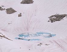
Watermelon snow, also called snow algaepink snowred snow, or blood snow, is Chlamydomonas nivalis, a species of green algae containing a secondary red carotenoid pigment(astaxanthin) in addition to chlorophyll. Unlike most species of fresh-water algae, it is cryophilic (cold-loving) and thrives in freezing water. Its specific epithet, nivalis, is from Latin and refers to snow.
This type of snow is common during the summer in alpine and coastal polar regions worldwide, such as the Sierra Nevada of California. Here, at altitudes of 10,000 to 12,000 feet (3,000–3,600 m), the temperature is cold throughout the year, and so the snow has lingered from winter storms. Compressing the snow by stepping on it or making snowballs leaves it looking red. Walking on watermelon snow often results in getting bright red soles and pinkish pant cuffs.

History

Watermelon snow streaks.
The first accounts of watermelon snow are in the writings of Aristotle. Watermelon snow has puzzled mountain climbers, explorers, and naturalists for thousands of years, some speculating that it was caused by mineral deposits or oxidation products that were leached from rocks.
In May 1818, four ships sailed from England to search for the Northwest Passage and chart the Arctic coastline of North America. Severe weather made them finally turn the ships back, but the expedition made valuable contributions to science. Captain John Ross noticed crimson snow that streaked the white cliffs like streams of blood as they were rounding Cape York on the northwest coast of Greenland. A landing party stopped and brought back samples to England. The Times wrote about this discovery on December 4, 1818.
A follow-up article three days later erroneously concluded that the coloration was caused by meteoric iron deposits.
When Ross published his account of the voyage in 1818, it contained a botanical appendix by Robert Brown. In it, Brown tentatively attributed the red snow to an alga.
The phenomenon was also reported from the Scottish Highlands in the nineteenth century and subsequently recorded scientifically from a snowpatch in the Cairngorm Mountains in 1967.[5]

Chlamydomonas nivalis

Unusual watermelon snow pits, superimposed with an orange-ish bootprint
Tracks made by sliding on watermelon snow in Utah's Uinta Mountains
Chlamydomonas nivalis is a green alga that owes its red color to a bright red carotenoid pigment, which protects the chloroplast from intense visible and also ultraviolet radiation, as well as absorbing heat, which provides the alga with liquid water as the snow melts around it. Algal blooms may extend to a depth of 25 cm (10 inches), with each cell measuring about 20 to 30 micrometers in diameter, about four times the diameter of a human red blood cell. It has been calculated that a teaspoon of melted snow contains a million or more cells. The algae sometimes accumulate in "sun cups", which are shallow depressions in the snow. The carotenoid pigment absorbs heat and as a result it deepens the sun cups, and accelerates the melting rate of glaciers and snowbanks.
During the winter months, when snow covers them, the algae become dormant. In spring, nutrients, increased levels of light and meltwater, stimulate germination. Once they germinate, the resting cells release smaller green flagellate cells which travel towards the surface of the snow. Once the flagellated cells reach the surface, they may lose their flagellae and form aplanospores, or thick-walled resting cells, or they may function as gametes, fusing in pairs to form zygotes.
Many species feed on C. nivalis, including protozoans such as ciliatesrotifersnematodesice worms and springtails.